Choroidal neovascular membrane (CNVM)

Choroidal neovascular membrane (CNVM)
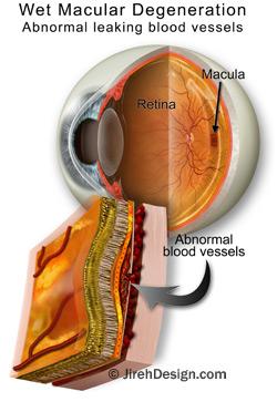
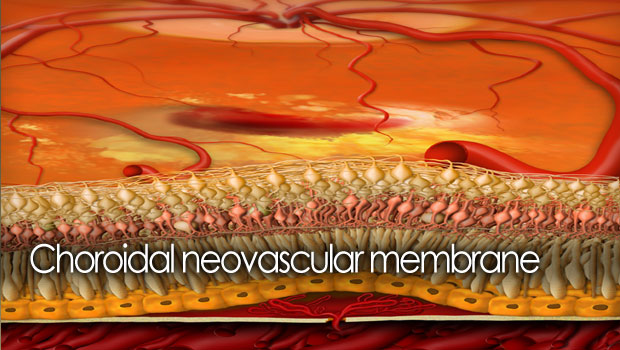
Choroidal neovascular membrane (CNVM), often seen in wet macular degeneration, involves the development of new, abnormal, leaking blood vessels in the retina.
CNVM is also called: Subretinal neovascular membrane (SRNVM), Choroidal neovascularization (CNV), Wet macular degeneration
What is CNVM?
Macular degeneration and other retinal diseases, like myopic degeneration and ocular histoplasmosis can damage the important layers of the retina. This tissue breakdown compromises the layer’s ability to act as a barrier to the vascular layer below the retina, called the choroid. This vascular layer is where a CNVM will typically develop.
Watch the “Human Eye” animation
When the retinal layers are damaged by diseases like macular degeneration, the choroid may produce new blood vessels (neovascularization) which grow up through the damaged layers and leak or bleed into the retina. Once this happens, the vision can become blurry, darkened or distorted. This form of AMD is called “wet macular degeneration”, as opposed to dry. See the differences between wet and dry macular degeneration here.
What are the symptoms of CNVM?
Since the retina acts as the “film in the camera” of the eye, any retinal irregularities in it can cause distortion, dark or fuzzy spots and loss of vision. If CNVM bleeds into the retina, the patient can experience dark, distorted or missing areas in the vision.
Using an Amsler grid daily is usually an effective way for the patient to detect any early changes in the central macular area — a common location of CNVM lesions.
How does my doctor diagnose a CNVM?
An ophthalmologist will usually diagnose a CNVM using in-office diagnostic tests such as Optical Coherence Tomography (OCT) or fluorescein angiography. Let’s take a look at both:
Fluorescein Angiogram
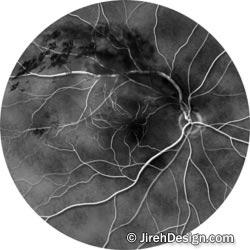
The fluorescein angiography findings also help the doctor choose which treatment is best for the particular patient and the photos will serve as documentation of the current state of the disease, allowing the doctor to follow and compare future study photographs. This series of dye tests will demonstrate the effectiveness of treatments and help the doctor decide if additional or alternative treatments should be done.
Optical Coherence Tomography (OCT) for macular degeneration
OCT is a non-invasive technology that is useful for imaging the retina, the multi-layered sensory tissue lining the back of the eye. This technique provides highly detailed, high-definition maps and images of the macula and retinal structures. Dr. Deupree can confirm the existence of CNVM and follow-up with this non-invasive study. This allows Dr. Deupree to make the most accurate diagnosis possible.
How do you treat CNVM?
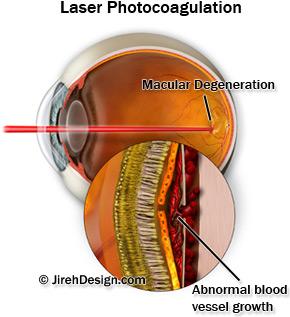
Of the two main forms of macular degeneration, wet and dry, wet macular degeneration is the only form with known, proven treatments. Those treatments include: Laser photocoagulation, Photodynamic Therapy, Macugen, Lucentis and Avastin injections.
Recently, the overwhelming treatment of choice throughout the vitreo-retinal medical community has been intravitreal anti-VEGF injections. These new medications target the underlying cause of CNVM in the “wet” form of macular degeneration. The abnormal blood vessels found in CNVM react to the medication by “drying up” after multiple injection applications. The results of these treatments has been very encouraging for macular degeneration patients.
See also…


