Vitreomacular Traction Syndrome
What is Vitreomacular Traction Syndrome? Vitreomacular traction (VMT) syndrome is an eye condition involving the vitreous, the clear gel that […]

Vitreomacular Traction Syndrome
What is Vitreomacular Traction Syndrome? Vitreomacular traction (VMT) syndrome is an eye condition involving the vitreous, the clear gel that […]
What is Vitreomacular Traction Syndrome?
Vitreomacular traction (VMT) syndrome is an eye condition involving the vitreous, the clear gel that fills the inside of the eyeball. It is a vision-threatening condition and can cause symptoms ranging from blurry vision to distorted and blacked-out central vision. Most patients who have VMT syndrome experience some difficulty doing daily tasks that require sharp vision, such as reading or watching TV.
Vitreomacular traction can lead to a macular hole. A macular hole will distort and blur the central vision and, in most cases, needs surgical intervention in order to reduce the likelihood of permanent vision loss.
To understand the mechanical causes of VMT, we should first familiarize ourselves with posterior vitreous detachment is and how it relates to VMT.
Posterior Vitreous Detachment (PVD)
The vitreous is a clear, gel-like substance (think of a jellyfish) that fills about 80 percent of the inside of the eyeball. The vitreous helps the eye maintain its round shape. There are millions of fine fibers intertwined within the vitreous that are attached to the surface of the retina – the eye’s light-sensitive tissue lining the back, inner wall of the eyeball. As the vitreous shrinks (usually due to aging) it becomes more liquefied and pockets of fluid form within the vitreous which leads to a contraction (syneresis) of the vitreous. Usually, not long after this process starts, the vitreous separates from the retina completely. This is called Posterior Vitreous Detachment (PVD).
Watch an animated vitreous detachment
PVD is a very common condition that usually affects people over age 50. Once the vitreous detaches, the condition is almost completely harmless.
What causes Vitreomacular Traction?
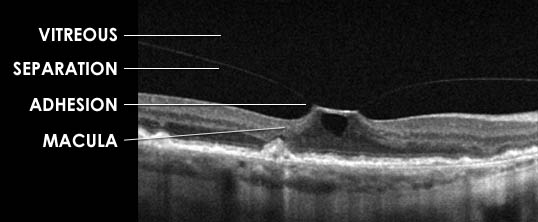
Vitreomacular traction occurs in cases where the vitreous adheres (vitreomacular adhesion) to the central vision part of the retina – the macula — instead of completely detaching. As only part of the vitreous attempts to pull away, a tugging or pulling occurs in the area of adhesion, causing actual physical alterations and distortion of the macula itself. (see diagram)
This persistent tugging on the macula usually causes swelling in its layers, a condition known as Cystoid Macular Edema (CME). CME can cause blurry vision and sometimes distortion of the vision.
How common is Vitreomacular Traction Syndrome?
VMT only occurs in about 1 in 4400 people. The occurrence of VMT in patients with diabetic retinopathy, age-related macular degeneration, and other macular diseases is much higher. It occurs in women slightly more often than men and can happen at any age, in any race.
How do you diagnose Vitreomacular Traction?
Optical Coherence Tomography (OCT) is a retina/vitreous scanner that provides an automated, segmented representation of the vitreous and macular layers. This non-invasive diagnostic study can show the retina specialist a cross-section view of the vitreous and macula. This image demonstrates the amount of involvement and tension on the macula caused by VMT. OCT is an extremely important tool in diagnosing and managing macular problems like VMT.
Fluorescein angiography is a valuable test that provides information about the condition of the retina. This dye study gives the eye doctor a “real-time” look at the circulation in the retina. It is also useful in finding any swelling, like CME, that may be present in the macula due to VMT.
 How is VMT treated?
Observation
Before the advent of Optical Coherence Tomography, doctors were under the impression that only 11 percent of eyes with VMT were naturally resolved by spontaneous detachment of the vitreous. Now that we have better diagnostic tools, it’s probably closer to 20 percent of VMT cases spontaneously resolve.
“Wait and see” is an option in some cases. However, symptoms must be closely observed with daily Amsler grid tests and follow-up visits to the ophthalmologist.
Surgery
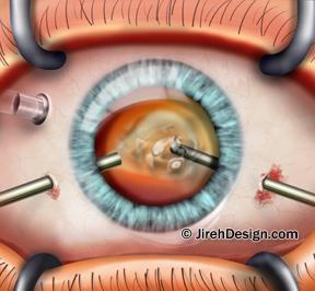
If VMT is threatening a macular hole, a pars plana vitrectomy is usually the best solution. This is a common retina surgical procedure involving removal of the vitreous. The retina specialist surgeon inserts a tiny, needle-like instrument, called a vitrector, into the eye. This instrument aspirates (sucks out) the vitreous fluid. The surgeon then refills the eye with a special saline solution that closely resembles the natural vitreous fluid in the eye.
The surgical removal of the vitreous relieves the traction on the macula and over time it will return to its normal structure and shape.
Ocriplasmin
Jetrea is an intravitreal eye-injected drug to treat Vitreomacular Adhesions. Jetrea is the first and only FDA-approved medicine for the treatment of VMA and it offers a much needed alternative to vitrectomy. The drug works by dissolving the protein fibers that create the adhesion between the vitreous and the macula. Dissolving this connection safely completes the detachment of the vitreous from the macula and relieves the traction.