Cirrus OCT – High Definition Retina Scanner
High definition OCT spectral domain retina scanner Dr. Deupree uses the newest, state-of-the-art, high-definition macula scanner on the market. The […]


Cirrus OCT – High Definition Retina Scanner
High definition OCT spectral domain retina scanner Dr. Deupree uses the newest, state-of-the-art, high-definition macula scanner on the market. The […]
High definition OCT spectral domain retina scanner
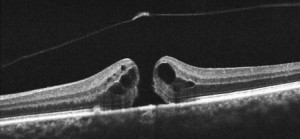
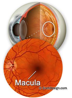
 Dr. Deupree uses the newest, state-of-the-art, high-definition macula scanner on the market. The Zeiss Cirrus HD-OCT is a non-invasive technology used for imaging the vitreous and retina — the multi-layered sensory tissue lining the back of the eye.
The Optical Coherence Tomography (OCT) scanner provides physicians with an automated, segmented representation of the choroid and retinal layers. It was the first instrument to allow doctors to see cross-sectional images of the retina. This technology has been used in vitreo-retina care for years. However, the latest high-definition, spectral domain scanning technology has revolutionized the way retina physicians image and diagnose macula conditions.

For macular degeneration, macular pucker, macular hole and glaucoma patients, this new imaging technology offers a quantum leap forward in diagnosis technology.


