Vitreous Hemorrhage
A patient’s complete, illustrated guide to vitreous hemorrhage The vitreous is normally a clear, jelly-like fluid that fills the inside […]


Vitreous Hemorrhage
A patient’s complete, illustrated guide to vitreous hemorrhage The vitreous is normally a clear, jelly-like fluid that fills the inside […]
A patient’s complete, illustrated guide to vitreous hemorrhage
The vitreous is normally a clear, jelly-like fluid that fills the inside of the eye. Various disease states can cause the vitreous to fill with blood so that light entering the eye will not reach the retina properly.
Also see “How Vision Works” animation
Vitreous Hemorrhage Definition
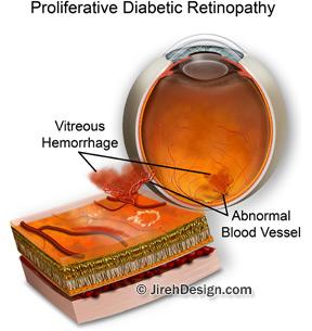
Vitreous hemorrhage, or bleed, results in a sudden change in vision as it blocks light moving through the vitreous to the retina. This hemorrhage specifically occurs in front of the retina in the posterior section of the eye.
The vitreous hemorrhage may be the result of an aneurysm of a blood vessel in the eye, trauma to the eye, a retinal tear, a retinal detachment, a new blood vessel (neovascularization) or as a result of another underlying disease state.
These disease states include diabetes, hypertension, sickle cell anemia, and carotid artery disease. Diabetics are particularly susceptible because the disease triggers the growth of new blood vessels within the eye.
The vessels are weak and bleed easily. This is why blindness is a concern for patients suffering from diabetes. Vitreous hemorrhage occurs more frequently in patients over 50 but can occur at any age.
Vitreous hemorrhage symptoms
Someone experiencing a vitreous hemorrhage may experience one or more of the following symptoms:
- sudden onset of blurry vision
- light flashes
- floaters (spots seemingly floating across the field of vision)
- blindness
Vitreous Hemorrhage Treatment
Initial treatment may be observation alone. Minor hemorrhages often clot and resolve on their own over time. Unfortunately, it may take months for full visual recovery from a vitreous hemorrhage.
Current research has produced drugs that can dissolve the vitreous gel inside the eye and may dramatically reduce the recovery time. These drugs are currently in the investigational stage and are awaiting FDA approval.
For more severe and debilitating vitreous hemorrhage, a vitrectomy may be performed. A vitrectomy is a surgical procedure that removes the vitreous gel and the blood from inside the eye.
After the vitreous is removed, the surgeon will refill the eye with a special saline solution that closely resembles the natural vitreous fluid in the eye.
Recovery from the procedure will take up to 6 weeks and complete vision recovery will take a little longer.


