Retinal artery occlusion


Retinal artery occlusion
What is the retina?
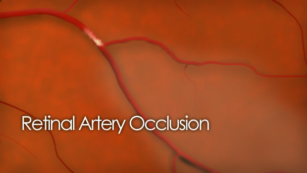
The retina, the light sensitive tissue that lines the back of the eye, is nourished by a fragile network of blood vessels including arteries and veins. The arteries carry important oxygen and nutrients to the eye where they are distributed out into the retinal tissues through a complicated meshwork of arterioles and capillaries. These nutrients are vital to the health and function of the retina. When an artery becomes blocked, it is termed a retinal artery occlusion.
Retinal arteries can become blocked by emboli, sometimes cholesterol or calcium plaques, that travel up the artery with the blood flow and become lodged where the artery or arteriole narrows. These emboli are often related to cardiovascular disease. Once this clogging occurs, the vital oxygen and nutrients are reduced or cut off from the retina that was supplied by the artery and the retina starves.
There are two types of retinal artery occlusions: branch retinal artery occlusion and central retinal artery occlusion.
Branch retinal artery occlusion (BRAO)
The smaller extensions off the main retinal artery are called “branches”. When a branch retinal artery becomes blocked, the retinal tissue that the branch artery supplies can be damaged, resulting in loss of vision. Edema and ischemia can occur and injury to the retina tissue is often permanent.
Typically, acute loss of vision is limited, to confined areas of the retinal and about 85% of eyes recover to 20/40 or better vision.
BRAO typically affects those over the age of 70. It is very rare in patients younger than 30, this is probably attributed to its link to cardiovascular disease.
Central retinal artery occlusion (CRAO)
The central retinal artery is the main blood supply to the retina. This main artery is actually fed by the ophthalmic artery which branches of off the carotid artery.
If an emboli lodges in the central retinal artery, the vision loss is profound in most cases. When obstruction occurs, it can take as little as 15 minutes for the retinal tissues to become ischemic and damaged.
Because the blockage takes place in the main artery that supplies blood to the retina (as opposed to a smaller branch artery), CRAO is more serious than BRAO. Vision loss usually ranges from as good as counting fingers to as bad as light perception only.
There is no well-proven treatment for central retinal artery occlusion with any high percentage of success. If the patient is in the doctor’s office quickly after the occlusion occurs, the doctor may attempt to massage the eyeball. This technique is often used to dislodge the embolus in hopes that it will travel further “downstream” in the blood vessel. The further downstream, the less of the retina that is affected, thus a smaller area of vision loss.

Some doctors also believe breathing into a paper bag can slightly dilate the retinal vasculature, promoting embolus movement.
None of the above-described procedures should be performed without the direction of a physician.


