Ophthalmoscopy eye examination
Ophthalmoscopy is a procedure that involves the examination of the back, inside part of the eye. It is usually performed […]

Ophthalmoscopy eye examination
Ophthalmoscopy is a procedure that involves the examination of the back, inside part of the eye. It is usually performed […]
Ophthalmoscopy is a procedure that involves the examination of the back, inside part of the eye.
It is usually performed by an eye doctor. Ophthalmoscopy, also known as fundoscopy, is a common procedure used to visualize the innermost structures of the eyeball, including the vitreous, retina, retinal blood vessels, macula and optic disc.
This short, 2-5 minute procedure is the cornerstone of a retina specialist’s examination process. It is used to detect eye diseases and conditions or to evaluate the patient’s symptoms. Conditions like retinal detachments and tears, macular pucker, diabetic retinopathy and glaucoma can be evaluated with this safe, non-invasive exam technique.
Ophthalmoscopy is also done on patients with systemic diseases that affect the blood vessels, like hypertension and diabetes. Ophthalmologists can often tell if a patient has a systemic disease just by looking at the retina using an ophthalmoscope.
 How is ophthalmoscopy performed?
Ophthalmoscopy requires that the pupil of the eye be dilated. This involves using a special eye drop formulated to enlarge the pupil opening and temporarily disable it from closing in response to bright light. A large, dilated pupil enables the doctor to see all the way out to the “peripheral” portions of the retina and vitreous. This is important, because many eye conditions begin in these outer lying areas of the retina.
There are three main types of ophthalmoscopy:
- Direct ophthalmoscopy: While the patient is seated in a dark exam room, the doctor uses a hand-held ophthalmoscope to shine a bright light into the eye. This instrument, about the size of a thin flashlight, uses many small lenses that focus on the inner structures of the eye and project the image back to the examiner.
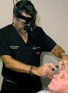
- Indirect ophthalmoscopy: This is usually done with the patient reclined back in the exam chair. The doctor holds the eye open while actually wearing the ophthalmoscope instrument on his/her head — think of a miner’s headlight. The doctor shines this headlight through a lens he’s holding in front of the eye. This lens magnifies the image that the doctor is seeing through the special mirrors and lenses attached to the ophthalmoscope. The patient is asked to look in various directions in order to move the area of interest into view.The doctor may employ a method called “scleral depression” where a slight pressure is applied to the side of the eye using a dull instrument. This is a technique that gives the doctor a better view of the far peripheral areas on the retina.
- Slit-lamp ophthalmoscopy: Probably the most commonly used type of ophthalmoscopy because the patient is usually already seated at a slit-lamp, an ophthalmic microscope that allows the doctor to view many areas of the eyeball at a various magnifications.
The patient sits upright in an exam chair in front of the slit-lamp with their chin rested on a support to keep their head steady. The doctor holds a tiny magnification lens in front of the eye and shines the slit-lamp light source through it. The enlarged image of the inside of the eye is reflected through the lens and seen by the doctor through the slit lamp binoculars. The doctor has the option to increase or decrease magnification using this method.
Does ophthalmoscopy hurt?
The bright light used during ophthalmoscopy can be uncomfortable for some. Unfortunately, it is necessary to illuminate the inside of the eye in order to get a detailed look. Though the brightness can even seem harmful, rest assured, it is a safe level of light that is being used.